
742: AI-3D Anatomical Mapping for Complex Robotic Partial Nephrectomy- Sanjoy Sureka
This is one of the entries accepted for adjudication for the 2026 KS Innovation Awards, sponsored by the Vattikuti Foundation. Posting here does not imply being a Finalist.
Producer: Dr. Sanjoy Sureka
Affiliation: Department of Urology and Renal Transplantation, Sanjay Gandhi Post Graduate Institute of Medical Sciences (SGPGI), Lucknow, India.
Email: drsksureka@gmail.com
Title: AI-Powered 3D Anatomical Mapping for Precision Robotic Partial Nephrectomy in Complex Renal Anatomy
Subtitle: For Management of Renal Tumors with Complex Anatomy
Purpose:
This video aims to demonstrate the performance of 3D Model in triaging 3D CECT KUB scans, specifically focusing on the detection, quantification, and localization of renal tumors in the setting of complex renal and vascular anatomy.
Materials and Methods:
3D Model utilizes advanced AI algorithms to detect, measure, and locate renal tumors, while also identifying their relation to aberrant vessels in cases of complex renal anatomy. Scans were processed using the AI algorithm, and all findings were carefully documented.
ITK Snap was employed for generating segmented scans, and tumor volume measurements were recorded. A radiologist with more than 5 years of dedicated urological imaging experience
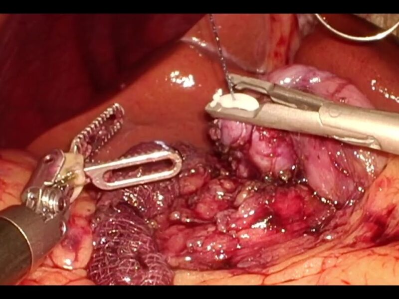
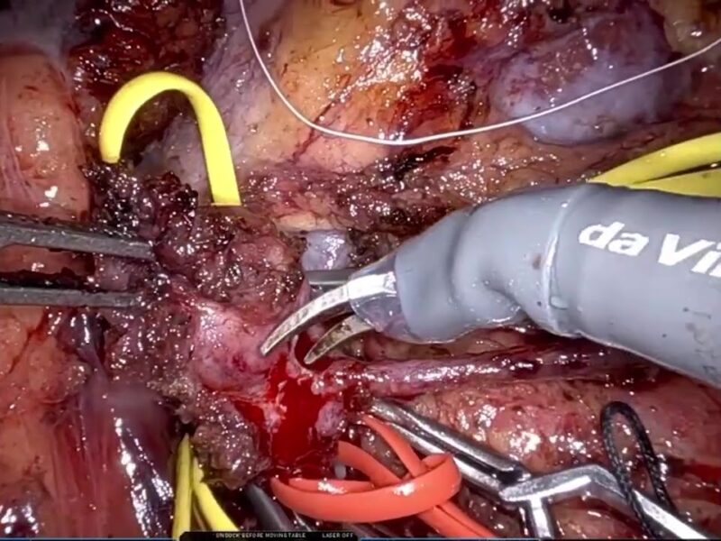
independently compared the AI-derived results with their own interpretations. The study highlights a 48-year-old male with a BMI of 42, presenting with a right ectopic pelvic kidney, aberrant arterial supply, and a renal mass. The patient subsequently underwent a robotic-assisted transperitoneal partial nephrectomy.
Results:
The 3D Model demonstrated sensitivity, specificity, and accuracy consistently exceeding 90% in detecting renal masses and their vascular supply. The operative duration was 118 minutes, with an
estimated blood loss of 85 cc and a warm ischemia time of 18 minutes. No major perioperative complications were encountered. The patient was ambulatory by postoperative day one and discharged home by day two. Histopathological analysis confirmed clear cell carcinoma grade 2 with negative surgical margins. Follow-up scans revealed no recurrence.
Conclusion:
The 3D Model proved highly effective in the triage of complex renal anatomy, offering reliable detection and accurate measurement of renal tumors, thereby contributing meaningfully to
patient management and precise surgical planning.
See more at: http://vattikutifoundation.com/
Date
August 15, 2020