3D Augmented Reality Guided Robotic-Assisted Kidney Transplantation
2A. Piana, 1A. Gallioli, 2D. Amparore, 1P. Diana, 1A. Territo, 3R. Campi, 1,2P. Verri, 1J.M. Gaya, 4L. Guirado, 2E. Checchucci, 2A. Bellin, 1A. Bravo, 1J. Palou, 3 S. Serni, 2F. Porpiglia, 1A. Breda Surgeon: 1Alberto Breda 1Department of Urology, Fundació Puigvert, Autonomous University of Barcelona, Spain 2Department of Urology, San Luigi Gonzaga Hospital, University of Turin, Italy 3Department of Urology, Careggi Hospital, University of Florence, Italy 4Department of Nephrology, Fundació Puigvert, Autonomous University of Barcelona, Spain
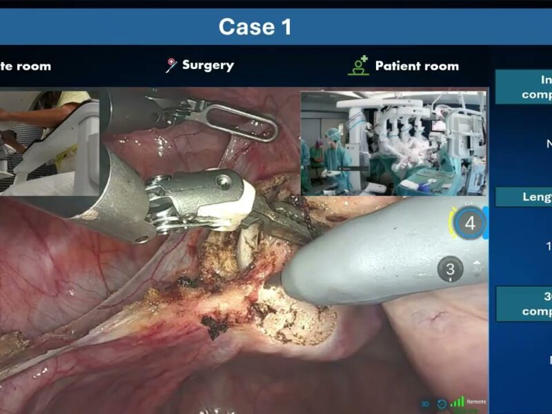
INTRODUCTION Robotic-assisted kidney transplantation (RAKT) has shown solid results as a minimally invasive alternative to the standard open approach (OKT) for living donor and promising results for deceased donor. However, this technique still holds a few limitations, such as its feasibility in case of atherosclerotic plaques in the recipient’s iliac vessels. During the robotic-assisted surgery, it is impossible to palpate the iliac plaques and thus to localize them. To overcome these limitations, we used three-dimensional (3D) virtual models in an augmented reality (AR) setting during RAKT to guide the surgeon during the clamping and arteriotomy phases.
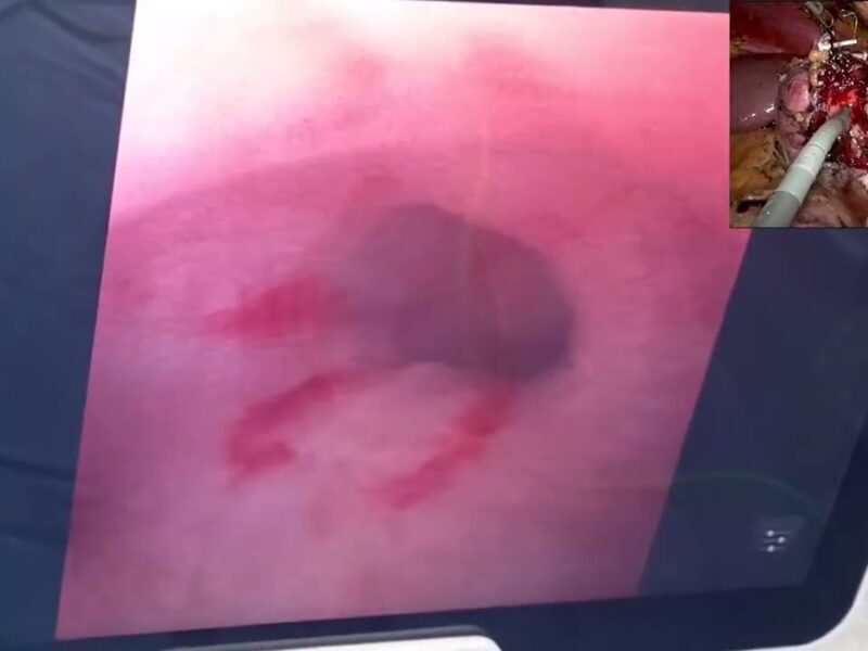
METHODS The study was conducted in accordance with the IDEAL model for surgical innovation. Initially (Phase 0), printed 3D-models were compared with the intraoperative anatomy during OKT. In Phase 1 a virtual model without plaques was superimposed on the real vessels during a combined open-robotic kidney transplantation and, later, during RAKT. Then, AR was employed during RAKT on a clinical case without plaques, during the clamping phase. Subsequently, this technology was tested on 10 patients with iliac atherosclerotic plaques during the lymph-node dissection’s phase of robot-assisted radical prostatectomy, verifying the correct position of the plaques using intraoperative ultrasound. Finally, the technology was applied during RAKT.
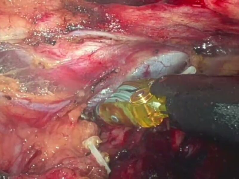
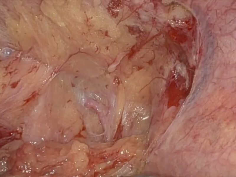
RESULTS AND CONCLUSIONS During RAKT, the 3D reconstructions allowed to avoid plaques, performing a successful clamping and a safe arteriotomy. The employment of 3D-AR allowed to overcome one of the main limitations of RAKT, setting the foundation to expand its indications to patients with advanced atheromatic vascular disease. Narrated Robotic Surgery video (3-D Augmented), with PPT’s, CT Scans 06:23
Date
August 15, 2020


