#353 Robot assisted 3D models-guided ureteral reimplantation- Dr.Federico Lavagno
This is one of the 2023 KS International Innovation Awards videos selected for inclusion in the Vattikuti Foundation – ORSI Humans at the Cutting Edge of Robotic Surgery Conference, October 6, 7 & 8, 2023 in Ghent, Belgium. Posting does not imply that is has been selected as a Finalist, just that the content will be discussed at the Conference.
From the entry: Robot assisted 3D models-guided ureteral reimplantation. Lavagno F, Allasia M, Oderda M, Marquis A, D’Agate D, Mangione C, Greco A, Pasquale G, Bosio A, Prof. Gontero P. Division of Urology, AOU Città della Salute e della Scienza di Torino, University of Turin School of Medicine.
INTRODUCTION AND OBJECTIVE The ureteral stenosis of the graft ureter is the most common urological complication of kidney transplants. In recurrent stenosis after stent removal, surgical revision is advocated.
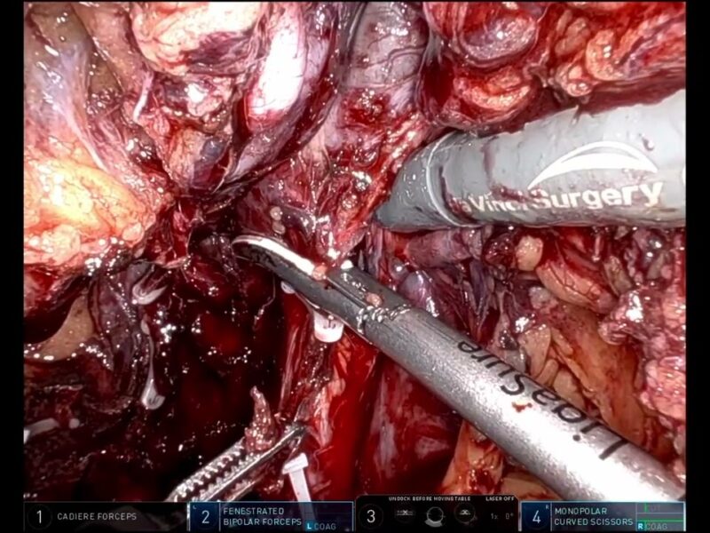
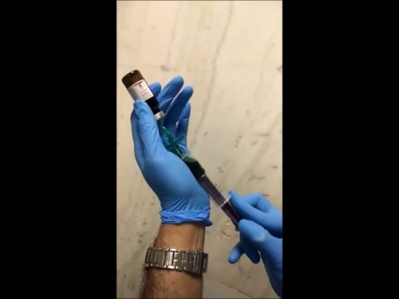
MATERIALS AND METHODS Patients with ureteral stenosis were treated with a Da Vinci robot with the use of pre operative 3D models and the auxilium of green indocyanine. Even if the scheduled intervention was the reimplantation of the ureteral graft to the bladder, this was not always possible. The key of the procedure is to identify and isolate the graft ureter transperitoneally. After the transverse opening of the isolated bladder it is made a uretero-vesical anastomosis. In other cases, instead, due to sclerotic tissue We harvested a Boari-Casati flap or We made a uretero-pyelo anastomosis between the native ureter and the graft kidney.
RESULTS To date, 7 patients (5 F and 2 M) with distal graft ureteral stenosis, planned for ureteral reimplantation. The mean time to onset of ureteral stricture was 3.5 months. Mean operative time was 217 minutes. Mean pre-op s-CR was 2.3 mg/dl, while post operative s-CR1.36 mg/dl. After 1 month observation mean s-CR was 1.45 mg/dl.
CONCLUSION The transperitoneal robotic approach with the use of 3D reconstructions and indocyanine could help in the identification and isolation of the ureter. 3D guidance can also make it easier to identify the renal vessels and the pelvis. On the other hand, the high costs of robotic surgery, further burdened by the use of 3D models, need to be considered.
See more at: https://vattikutifoundation.com/videos/
Date
August 15, 2020


