585 Surgical Nuances of Robotic Reconstruction in Genito-urinary Tuberculosis
Dr. Rohit Deshpande
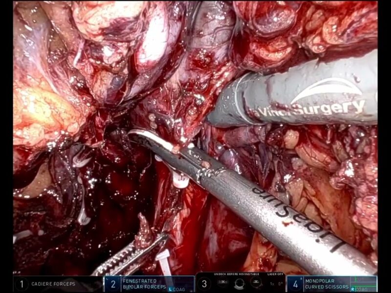
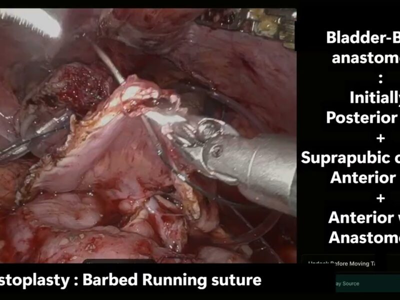
A young lady presented to us with debilitating flank pain, subsequent to scarring and stricturing of the left pelvi-ureteric junction, which was sited intra-renally. She had a past history of being treated for genitourinary tuberculosis, and had completed anti-tuberculous therapy. On imaging, she had irregular caliectasis of the left kidney, which typified the tuberculous etiology, and presented to us a complex problem. 3-dimensional computed axial imaging was done and subsequently she underwent a robotic reconstruction of the left kidney, necessitating a uretero-calycostomy. The video highlights the dense periureteric and peri-pelvic fibrosis associated with the tuberculous stricture. Also, intra-operative endoscopy was done to inspect the pelvicalyceal system for a renal stone (which was sited in a calyceal diverticulum). Post-operatively, the half-clearance time of the radiotracer on renal scintiscan had reduced significantly (thus proving un-obstructed drainage), with rapid symptomatic resolution.
See more at: https://vattikutifoundation.com/videos/
Date
August 15, 2020