Robot assisted Nephroureterectomy for Transitional cell carcinoma in a horseshoe kidney
This video was an entry for the 2020 KS National Robotic Surgery Video Awards: Robot assisted Nephroureterectomy for Transitional cell carcinoma in a horseshoe kidney; Amit Bansal, Ruchir Maheshwari, Samit Chaturvedi, Devanshu Bansal, Anant Kumar Department of Urology, Renal Transplantation, Robotics and Uro-oncology; Max Super-specialty Hospital, New Delhi, India
Abstract: Introduction: We present, and discuss the operative steps, mistakes made, and the important lessons learnt during robot-assisted radical nephroureterectomy with bladder cuff excision, performed for transitional cell carcinoma in a patient with horseshoe kidney.
Patient and method: Seventy-four-year-old gentleman with the history of recurrent hematuria with passage of clots, since last one year, presented with a CT scan, suggestive of mass in the right renal pelvis, with horseshoe configuration of the kidneys. His urine cytology was negative for malignant cells. He underwent flexible ureteroscopy and biopsy from the mass, which was suggestive of low-grade transitional cell carcinoma. Later he underwent robot-assisted right nephroureterectomy with bladder cuff excision.
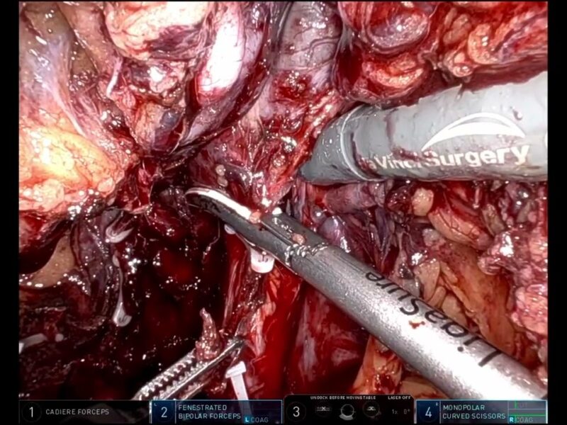
Results: Cystoscopy done at the time of flexible ureteroscopy did not reveal any tumor in the urinary bladder. He later underwent robot-assisted right nephroureterectomy with bladder cuff excision. The patient was placed in lateral position with the right side up. Ports were placed in an oblique fashion. Salient steps included, the configuration of ports, colon mobilization and hilar dissection, vascular dissection, division of isthmus and hemostasis, mobilization of the ureter and bladder cuff excision. We present the aberrant vasculature encountered intra-operatively, despite the previous discussion with a radiologist, and the management of the same. We noticed duodenal injury intra-operatively and managed it with primary repair. We further discuss the steps of isthumectomy and bleeding control.
Conclusions: Key takeaway points from this presentation are emphasis on pre-operative discussion of CT with the radiologist to better delineate vascular anatomy. However, we may still encounter any intra-operative surprise. Patient and meticulous dissection at the hilum, helped us to identify previously missed major renal vessels. One must stay alert to detect any injury intra-operatively and promptly repair the same. Identification of a line of demarcation over the isthmus, helps to identify the correct area to divide the isthmus, and minimize the bleeding. Optimal use of versatility of da Vinci Xi system, enables the port placement and switching the robotic instruments and camera between the four ports, according to the comfort of the operating surgeon.
Narrated robotic surgery video, with CT, clinical and operative photographs, diagrams and robotic surgery clips, duration – 07:51
Date
August 15, 2020


