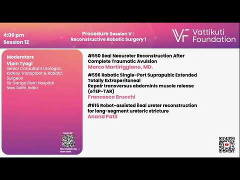
#241: Improving margin assessment during prostatectomy with ex-vivo FCM Dr. Ricardo Almeida-Magana
This is one of the 2023 KS International Innovation Awards videos selected for inclusion in the Vattikuti Foundation – ORSI Humans on the Cutting Edge of Robotic Surgery Conference, October 6, 7 & 8, 2023 in Ghent, Belgium. Posting does not imply that is has been selected as a Finalist, just that the content will be discussed at the Conference.
From the entry: We describe a technique to analyse surgical margins during robotic radical prostatectomy using a fluorescence confocal microscope and report its accuracy metrics.
ABSTRACT: Improving margin assessment during prostatectomy with ex-vivo FCM Ricardo Almeida-Magana University College London Hospital United Kingdom
Introduction Frozen section analysis (NeuroSAFE) during robot-assisted radical prostatectomy (RARP) is accurate but time-consuming and expensive. Ex-vivo fluorescence confocal microscopy (FCM) offers a promising alternative with minimal tissue processing. However, previous studies lacked sufficient positive surgical margins (PSM) cases for accurate assessment. This video presents a novel FCM method for evaluating RARP specimens and reports its accuracy.
Methods Patients undergoing non-nerve-sparing RARP for suspected extraprostatic extension were included. After extraction, the specimen was treated with a photoreactive dye, rinsed, and examined using the Histolog scanner®, an FCM-based microscope. A high-resolution digital image of the specimen’s posterolateral surface was stored for later review. No clinical decisions were based on FCM results, and the specimen was reattached for final paraffin analysis. Informed consent was obtained from all patients.
Results Thirty-one specimens were analyzed, with a median tumour volume of 3.25 mL. Most specimens were graded as Gleason 3+4. Two independent uro-oncology histopathologists reviewed 59 good- quality FCM images, unaware of the paraffin analysis results. Sensitivity and specificity were calculated. The first reviewer achieved 88.9% sensitivity and 94.1% specificity. The second reviewer achieved 66.7% sensitivity and 96.1% specificity. No subsequent tissue processing was affected.
Conclusions This proof-of-concept study demonstrates the accuracy of ex-vivo FCM imaging for diagnosing PSM during RARP. Further research is needed to compare its performance to NeuroSAFE and evaluate its clinical usefulness in larger patient cohorts.
See more at: https://vattikutifoundation.com/videos/
Date
August 15, 2020