Confocal Microscopy for Surgical Margins During Prostatectomy
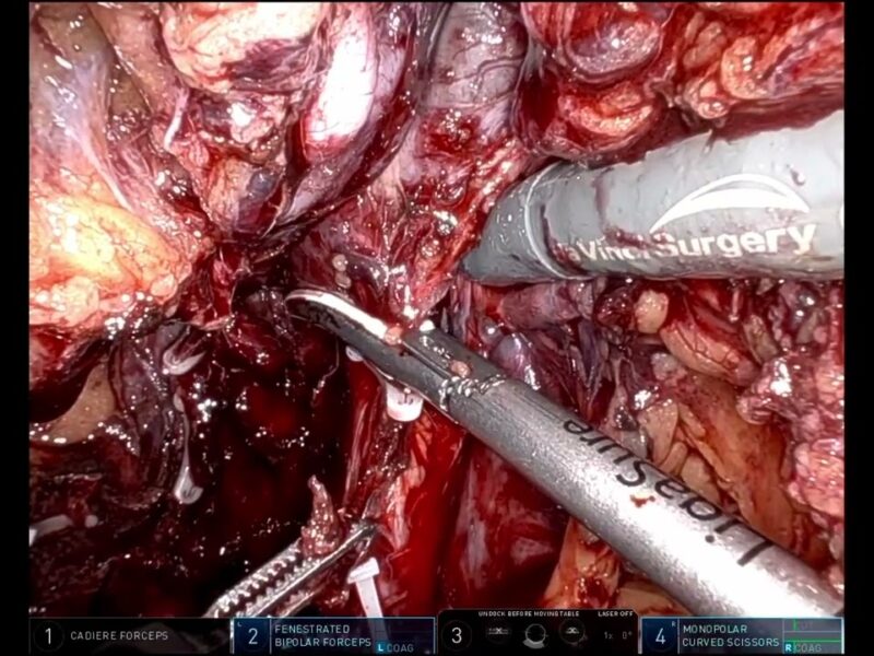
This video was entered by Dr. Paolo Verri in the 2022 KS International Robotic Surgery Innovation Awards, sponsored by the Vattikuti Foundation. It was featured in the Vattikuti Symposium ‘Humans at the Cutting Edge of Robotic Surgery,’ held in Miami, Florida November 19, 2022.
Here is the Abstract:
CONFOCAL MICROSCOPY FOR SURGICAL MARGINS DURING RADICAL PROSTATECTOMY
P. Verri, A. Gallioli, P. Diana, A. Territo, F. Sanguedolce, I. Sanz, M. Baboudjian, J. Huguet, J.M. Gaya, F. Algaba, J. Palou, A. Breda
Surgeon: Alberto Breda
Department of Urology, Fundació Puigvert, Autonomous University of Barcelona, Spain
Introduction: During radical prostatectomy (RP), a nerve sparing approach can be performed to improve postoperative functional outcomes, although the risk of positive surgical margins can increase. In this study, we aim to validate the reliability of confocal microscopy in the diagnosis of positive surgical margins during minimally invasive RP.
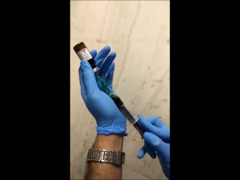
Methods: VivaScope® 2500M-G4 microscope (Mavig GmbH, Munich, Germany; Caliber I.D.; Rochester NY, USA) is a laser scanning microscope, capable of obtaining high quality images with a thickness of less than 5.0μm thanks to a laser beam with different wavelengths. In this ongoing single-center prospective study, approved by the Ethical Committee, all patients undergoing minimally invasive RP are enrolled. After the surgical procedure, two tissue specimens are harvested from the prostatic postero-lateral margins and analyzed with CM before sending them to final pathology.
Results: On July 1st 2022, 30 patients (60 surgical margins) had been enrolled. According to d’Amico classification, 2 (6.7%), 16 (53.3%) and 12 (40%) patients had low-, intermediate- and high-risk disease, respectively. At MRI, 5 (16.6%) and 3 (10%) patients had extracapsular extension and seminal vesicles invasion. At final pathology, there were 10/30 (30%) positive surgical margins. Intraoperative diagnosis with CM was obtained in 6 (4-7) minutes. After CM, 8/60 (13.3%) margins were positive. CM showed a sensitivity (95% CI), specificity, NPV, and PPV of 0.7 (0.35-0.91), 0.98 (0.87- 0.99), 0.94 (0.83-0.98) and 0.87 (39.4-90.5), respectively.
Conclusions: Ex-vivo CM during RP provides a fast and reliable intraoperative analysis of prostatic surgical margins.
See more at: vattikutifoundation.com/videos
Date
August 15, 2020


