#226 D-MODELS GUIDANCE FOR ROBOTIC RENAL ARTERY ANEURYSM REMOVAL- Dr. Daniele Amparore
This is one of the 2023 KS International Innovation Awards videos selected for inclusion in the Vattikuti Foundation – ORSI Humans on the Cutting Edge of Robotic Surgery Conference, October 6, 7 & 8, 2023 in Ghent, Belgium. Posting does not imply that is has been selected as a Finalist, just that the content will be discussed at the Conference.
From the entry: 3D-MODELS GUIDANCE FOR ROBOTIC RENAL ARTERY ANEURYSM REMOVAL Daniele Amparore, Federico Piramide, Paolo Verri, Simona Barbuto, Enrico Checcucci, Sabrina De Cillis, Alberto Piana, Mariano Burgio, Giovanni Busacca, Marco Colombo, Cris@an Fiori, Francesco Porpiglia San Luigi Gonzaga Hospital – Candiolo Cancer Institute, University of Turin (Italy)
Introduction Recently, 3D virtual models (3DVMs) have paved the way towards the knowledge of surgical anatomy. Their newest generation can identify arterial perfusion volumes within the organ, finding a role in the surgical management of renal artery aneurysms. We present the case of a renal artery aneurysm, underwent 3DVM-guided robotic surgery.
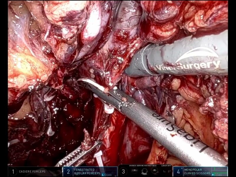
Methods As assessed with the 3DVM the pre-pyelic branch of the right renal artery presented an aneurysm and a shunt with a vein. Five kidney perfusion volumes and three vascular management strategies were identified: – to clamp the entire pre-pyelic branch, excluding the three perfusion volumes of the lower half of the kidney; – to clamp the third-order aneurysmatic pre-pyelic branch before its division in fourth order arteries, devascularizing two mesorenal perfusion volumes, but sparing the lower pole of the organ – to clamp the aforementioned artery, downstream of the fourth-order branch, devascularizing only the volume of kidney fed by the aneurysmatic vessel. The surgical strategy then involved the arteriovenous shunt closure and the removal of the aneurysm, preserving the kidney.
Results IntroperaAvely, the pre-pyelic branch was clamped upstream of the aneurysm, as suggested by second opAon planned with 3DVM, and the shunt clipped. Additional clip was put on the prepyelic artery below its division in fourth-order branches, following the third 3DVM-based planning strategy. No perioperative complications were recorded, while the renal funcAon was conserved postoperatively.
Conclusions The planning and surgical management of complex vascular anatomies with new-generation 3DVMs leads to minimize intraoperative risks while maximizing the functional results.
See more at: https://vattikutifoundation.com/videos/
Date
August 15, 2020


